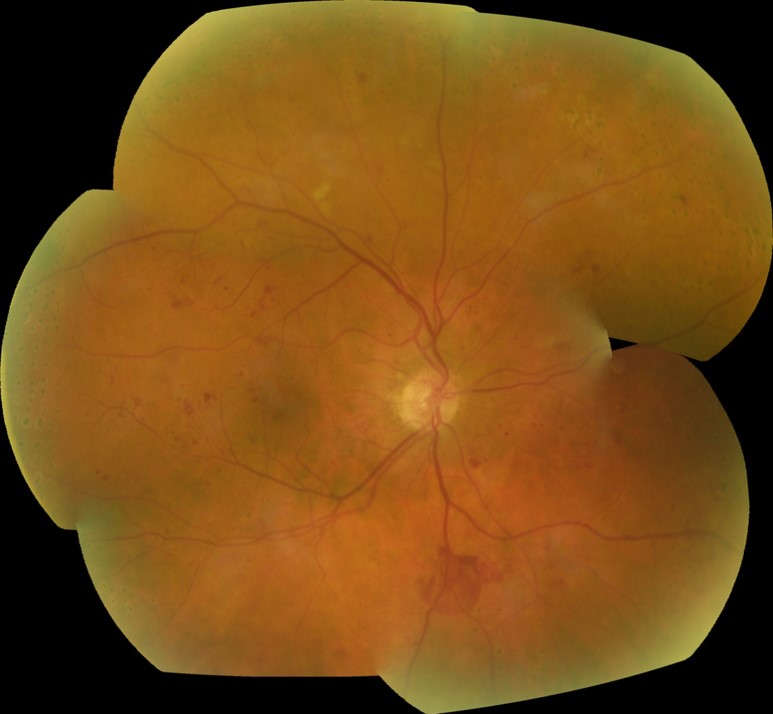
7 Modified Field Capture
Capture Tutorial: A tutorial showing the following 7 modified field capture, with examples, is available for download here.
Capture Video:In addition, this video shows the capture of 7 fields on a Spectralis system.
| Field 1M - Optic disc: | Asking the patient to look at the external fixator, centre the temporal edge of the optic disc in the field. If using a fundus camera, this will be at the intersection of the ocular crosshairs. |
| Field 2 - Macula: | Simply pivot the camera towards the temporal area from F1 without any vertical adjustment. The fovea should be positioned just below the centre of the field. This helps to avoid possible central grey artefacts created by some cameras. |
| Field 3 - Temporal to macula: | Pivot the camera further toward the temporal area to position the macula mid way between the centre of the field and the nasal edge of the field, again without vertical adjustment. You may need to use the internal fixator to achieve this. |
| Field 4 - Superior temporal: | Centre the optic disc in the frame, tilt the camera downwards until the optic disc is at the 6 o’clock position, pivot the camera towards the temporal area to position the disc in the lower right corner of the field. If using a 30 or 35° lens, position the disc just outside the frame, making sure that if it’s not all visible, that three closely arranged retinal vessels are showing. |
| Field 5 - Inferior temporal: | Centre the optic disc in the frame, tilt the camera upwards until the optic disc is at the 12 o’clock position, pivot the camera towards the temporal area to position the disc in the upper right corner of the field. If using a 30 or 35° lens, position the disc just outside the frame, making sure that if it’s not all visible, that three closely arranged retinal vessels are showing. |
| Field 6 - Superior nasal: | Centre the optic disc in the frame, tilt the camera downwards until the optic disc is at the 6 o’clock position, pivot the camera towards the nasal area to position the disc in the lower left corner of the field. If using a 30 or 35° lens, position the disc just outside the frame, making sure that if it’s not all visible, that three closely arranged retinal vessels are showing. |
| Field 7 - Inferior nasal: | Centre the optic disc in the frame, tilt the camera upwards until the optic disc is at the 12 o’clock position, pivot the camera towards the nasal area to position the disc in the upper left corner of the field. If using a 30 or 35° lens, position the disc just outside the frame, making sure that if it’s not all visible, that three closely arranged retinal vessels are showing. |